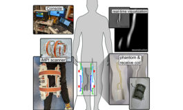
Researchers at the University of Würzburg in Germany have developed a portable scanner that is the first to be able to image humans using Magnetic Particle Imaging (MPI). The technology could provide a radiation-free alternative to techniques...
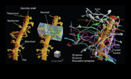
Researchers at the Institute of Science and Technology Austria have developed a brain imaging technique called Live Information Optimized Nanoscopy Enabling Saturated Segmentation (LIONESS). The method lets the researchers create...
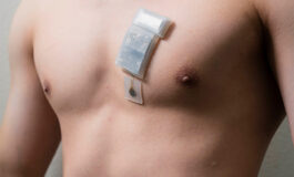
Researchers at the University of California San Diego have created a wearable ultrasound system that can monitor deep tissues, as far as 16.5 cm (6.5 inches) below the surface of the body. Moreover, the team employed a machine learning...
Researchers at Northwestern University have trailed an implanted ultrasound device in patients, which is used in combination with microbubbles to transiently open pores in the blood brain barrier, allowing chemo drugs to enter. We have reported...
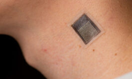
Researchers at the University of California San Diego have developed a wearable ultrasound patch that is intended to provide information on the stiffness of underlying tissues as deep as 4 cm below the surface of the skin. The patch consists of...
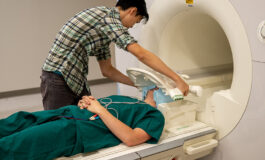
Researcher scientists at the University of Texas at Austin have developed a brain decoding technology that combines an fMRI scanner and artificial intelligence, similar to well-known AI systems such ChatGPT or Bard. The technology can spell out...
RF (radio frequency) technology uses radio waves to transmit and receive information wirelessly. RF is often seen in health monitoring devices such as wearables, implants, remote monitoring systems, and telemedicine. Mostly, though, it is used...
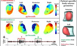
Researchers at the Eunice Kennedy Shriver National Institute of Child Health and Human Development have developed a new imaging technique called electromyometrial imaging (EMMI) which clinicians can use to create 3D maps of uterine contraction...
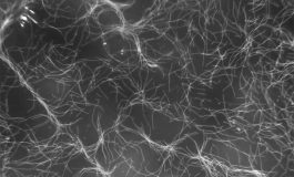
Researchers at Caltech have developed a technique that lets them move groups of cells very precisely. It involves genetically modifying cells so that they express small protein air sacs in their interior. The sacs render the cells highly...
Researchers at Columbia University Irving Medical Center and the Université de Paris, France, have tested an ultrasound denervation catheter in its potential to treat hypertension. The technology is called the Paradise ultrasound denervation...
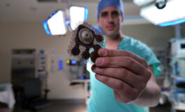
Researchers at RMIT University in Australia have developed ‘smart stitches’ that can fight bacteria and reveal the location of the sutured area in CT scans. The sutures have been developed to reduce the chances of surgical site infections...